Introduction
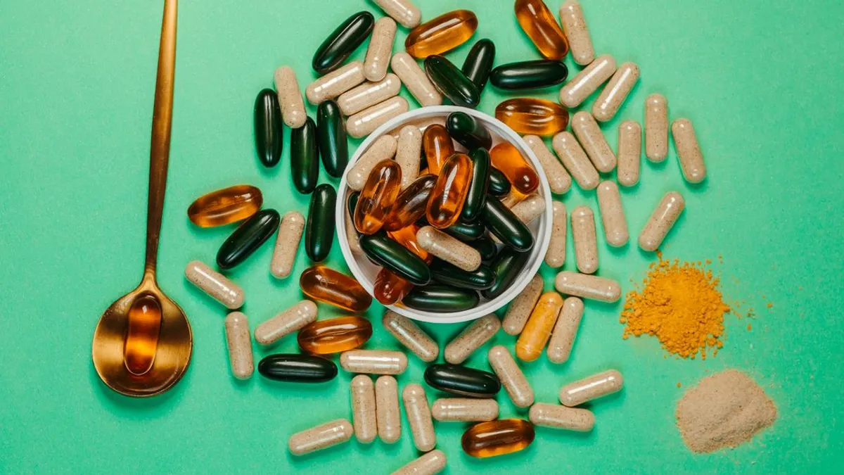
Vitamin D is the most prescribed nutraceutical in endocrinology — almost every patient with autoimmune thyroiditis (AIT), chronic fatigue, or metabolic syndrome has their 25(OH)D measured. Yet the clinical effect of isolated D3 therapy is often weaker than expected, and patients on high doses develop side effects ranging from hypercalcemia to progressive coronary artery calcification. The reason: D3 solves calcium absorption, but not calcium distribution. Distribution is governed by vitamin K2.
This article covers the biological pair D3 + K2 (menaquinone MK-7) in the context of Hashimoto autoimmune thyroiditis. Why one without the other works only half-way or even harms, what the 25(OH)D targets are in AIT, how D3 affects thyroid peroxidase antibodies (TPO-Ab), which cofactors are mandatory, and who must avoid the pair. Recommendations follow the Endocrine Society guideline, meta-analyses, and controlled RCTs.
🌀
What D3 and K2 measure: distinct biochemistry, shared target
Vitamin D3 (cholecalciferol) is synthesized in skin under UVB radiation from 7-dehydrocholesterol and enters with food (fatty fish, egg yolk, liver). In the liver it is hydroxylated to 25(OH)D (calcidiol) — the serum metabolite measured as D status. In the kidney, 25(OH)D is converted by 1α-hydroxylase into the active 1,25(OH)₂D (calcitriol), which binds the nuclear VDR and regulates expression of more than 200 genes, including calcium-binding proteins of intestinal epithelium. Result: dietary calcium absorption rises from 10–15% to 30–40%.
Vitamin K2 (menaquinone, MK) is a fat-soluble cofactor of γ-glutamyl carboxylase, the enzyme that converts inactive Gla proteins (containing glutamate) into active calcium-binding forms. Two Gla proteins are clinically pivotal:
▸Osteocalcin (OC) — produced by osteoblasts; in its carboxylated form it binds Ca²⁺ and embeds it into bone hydroxyapatite. ▸Matrix Gla protein (MGP) — produced by vascular smooth muscle cells; in its carboxylated form it blocks Ca²⁺ deposition on elastin fibers of the arterial wall.
Without sufficient K2 these proteins remain undercarboxylated (ucOC and dp-ucMGP) — calcium mobilized by D3 from the gut neither enters bone nor is blocked from vessels. Schwalfenberg (PMID 28698808) showed that patients on high-dose D3 (≥5000 IU/day) without K2 have increased vascular calcification risk and reduced insulin sensitivity.
MK-7 is the best-studied and pharmacologically favourable K2 form: its half-life is substantially longer than that of MK-4 (Sato et al., PMID 32244313). This means 100–200 µg of MK-7 once daily delivers stable γ-carboxylation, while MK-4 requires dosing three times a day.
🌀
Clinical: 25(OH)D and autoimmune thyroiditis
The link between vitamin D deficiency and autoimmune thyroiditis is confirmed in dozens of observational studies and several RCTs. A meta-analysis by Wang et al. (PMID 29388046) pooled 6 RCTs (n = 344) and showed that adding D3 significantly reduces TPO-Ab over 6 months versus control. The effect is statistically significant (p < 0.01) and clinically reproducible.
Target 25(OH)D levels remain debated. The Endocrine Society guideline (Holick et al., PMID 21646368) defines deficiency as < 20 ng/mL (< 50 nmol/L), sufficiency as > 30 ng/mL. For AIT patients the optimal target is 40–60 ng/mL (100–150 nmol/L) — the range where RCTs show maximal reduction of TPO-Ab and improved T4 → T3 conversion while preserving safety (no hypercalcemia).
The molecular mechanism of D3 on thyroid function is multilevel. (1) VDR is expressed on regulatory T cells (Treg); active 1,25(OH)₂D increases their count and function, restoring the Th17/Treg balance disturbed in Hashimoto. (2) D3 lowers autoantigen presentation by dendritic cells. (3) IFN-γ production by Th1 cells and IL-17 by Th17 cells — the two central cytokines of thyrocyte autoimmune destruction — are suppressed. (4) Deiodinase activity improves indirectly through reduced systemic inflammation, important for functional hypothyroidism (see T4 → T3 conversion and deiodinases).
Important: D3 + K2 do not replace levothyroxine in overt hypothyroidism. If TSH > 4.5 mIU/L with low free T4 or classic hypothyroid symptoms, the base remains replacement therapy (levothyroxine or Thyroid-S NDT — see hypothyroidism and natural desiccated thyroid NDT). D3 + K2 is a nutritional superstructure that reduces autoimmune activity and supports bone-vascular health on top of primary therapy.
🌀
Common pitfalls
Most errors in D3 prescribing are not about dose but about what falls out of view.
▸K2 is not prescribed at all. Standard practice gives a patient with D3 deficiency 5000–10000 IU/day for 2–3 months without K2. Calcium is mobilized from gut, but without active osteocalcin and MGP it deposits in vessels. Per Schwalfenberg (PMID 28698808), high-dose D3 without K2 raises dp-ucMGP — a biomarker of arterial calcification. ▸Magnesium is ignored. Magnesium is a cofactor of every D-metabolism enzyme: CYP2R1 (hepatic hydroxylase), CYP27B1 (renal 1α-hydroxylase), CYP24A1 (catabolism). With Mg²⁺ deficiency (serum Mg²⁺ < 0.75 mmol/L or RBC-Mg²⁺ < 4.2 mg/dL), even large D3 doses fail to raise 25(OH)D — the vitamin stays "asleep". ▸Albumin-corrected calcium is not monitored. On high-dose D3 (≥5000 IU/day) total and ionized calcium must be measured every 8 weeks. Hypercalcemia is rare but dangerous. ▸Interaction with thyroxine is overlooked. High-dose D3 may accelerate levothyroxine metabolism by induction of hepatic CYP — some patients require dose adjustment after 8–12 weeks. ▸K2 is prescribed to warfarin patients. Menadione (K3) and MK-7 neutralize the warfarin effect — INR drops sharply, thrombotic risk rises.
🌀
Protocol: doses and timing
Doses depend on baseline 25(OH)D. AIT target range: 40–60 ng/mL.
| 25(OH)D | D3 dose |
|---|---|
| < 20 ng/mL | 5000 IU/day × 8 wk |
| 20–30 ng/mL | 2000–4000 IU/day × 8 wk |
| 30–40 ng/mL | 1000–2000 IU/day |
| 40–60 ng/mL | 1000 IU/day maintenance |
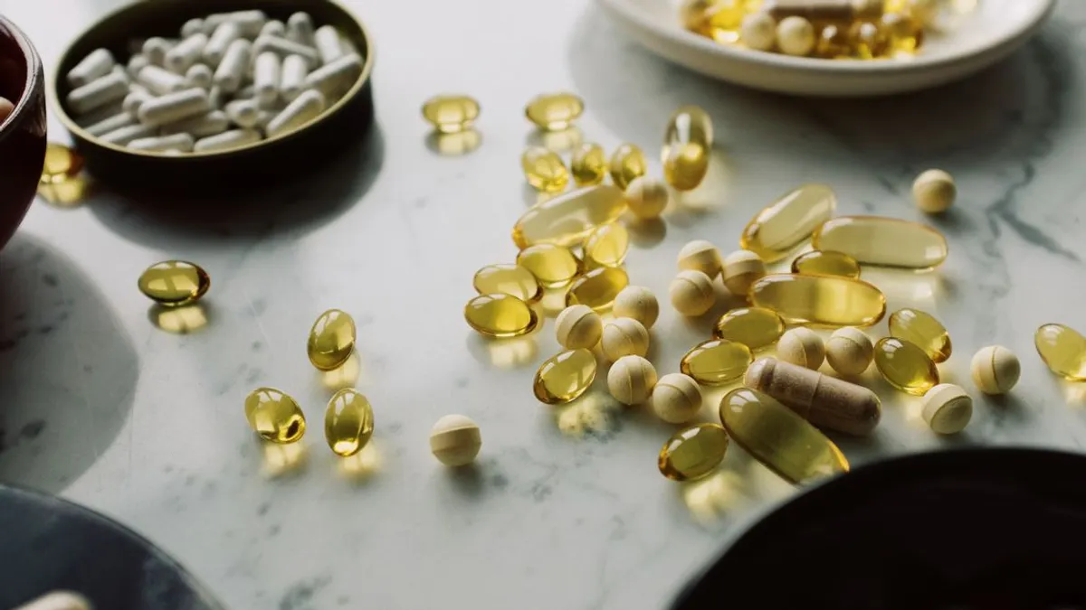
▸Deficiency, 25(OH)D < 20 ng/mL (< 50 nmol/L) — D3 5000 IU/day × 8 weeks, then 2000 IU/day maintenance. ▸Insufficiency, 25(OH)D 20–30 ng/mL (50–75 nmol/L) — D3 2000–4000 IU/day × 8 weeks, then 2000 IU/day. ▸Sufficient, 25(OH)D 30–40 ng/mL — D3 1000–2000 IU/day to reach the 40–60 ng/mL target. ▸Optimal, 25(OH)D 40–60 ng/mL — D3 1000 IU/day maintenance + seasonal correction (+500 IU in winter). ▸K2 (MK-7) — 100 µg/day with D3 ≤2000 IU/day; 200 µg/day with D3 ≥4000 IU/day. Doses above 360 µg/day do not increase γ-carboxylation. ▸Magnesium — 200–400 mg of elemental magnesium as glycinate, citrate, or threonate (NOT oxide — bioavailability < 5%). Recheck RBC-Mg²⁺ at 8 weeks. ▸Vitamin A (retinol) — 1000–3000 IU/day as retinyl palmitate or from cod liver oil. Not β-carotene in thyroiditis — conversion to active retinol is reduced. ▸Boron — 3 mg/day (optional cofactor that potentiates bone-density effect). ▸Administration — all together with a fat-containing meal (egg yolk, avocado, nuts, olive oil). D3 and K2 are both fat-soluble; absorption with water is ≤20%.
Do not separate D3 and K2 in time. Modern combination products (D3 5000 IU + K2 100 µg in one capsule) are the optimal delivery form. Splitting the two more than 4 hours apart reduces Gla-protein synergy.
🌀
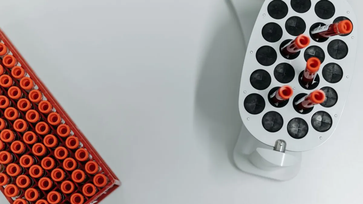
Monitoring
Minimum laboratory monitoring on D3 + K2:
▸25(OH)D — baseline, 8 weeks after start/dose change, then every 6 months on maintenance. ▸Albumin-corrected Ca²⁺ — baseline and at 8 weeks on doses ≥4000 IU/day. ▸PTH (parathyroid hormone) — baseline if secondary hyperparathyroidism is suspected (often with D < 15 ng/mL). ▸RBC-Mg²⁺ or serum Mg²⁺ — baseline and at 8 weeks. ▸TPO-Ab, TSH, free T3, free T4 — baseline and at 6 months for autoimmune-process effect assessment. ▸Osteocalcin (carboxylated + ucOC) — optional but informative when AIT coexists with osteopenia/osteoporosis. ▸Plasma K2 is not measured routinely — assessment goes through indirect markers: cOC/ucOC ratio, dp-ucMGP, bone mineral density (DEXA after 1 year).
If 25(OH)D fails to rise as expected at 8 weeks (< +10 ng/mL) — check magnesium, rule out malabsorption (celiac, SIBO, post-bariatric syndrome), revisit the dose.
🌀
Caution: contraindications and interactions
D3 + K2 is therapy with real pharmacological action and carries strict contraindications.
▸Warfarin and other vitamin K antagonists — K2 neutralizes the anticoagulant effect. Absolute contraindication without cardiologist sign-off. If K2 is essential (severe osteoporosis), transitioning to a DOAC (apixaban, rivaroxaban) is discussed with cardiology. ▸Hypercalcemia of any cause — stop D3 until the cause is addressed (primary hyperparathyroidism, myeloma, metastases, prior D overdose). ▸Sarcoidosis, lymphoma, granulomatous disease — granuloma macrophages produce 1,25(OH)₂D endogenously from 25(OH)D, bypassing renal control. Even physiologic D3 doses can cause hypercalcemia. D3 dose is minimal (≤1000 IU/day) under Ca²⁺ control. ▸Severe chronic kidney disease (CKD 4–5) — 1α-hydroxylase activity is reduced; active forms (alfacalcidol, calcitriol) are prescribed under nephrology supervision, not native D3. ▸Active nephrolithiasis (calcium oxalate stones) — increased calcium excretion on D3 therapy may aggravate stone formation; assess 24-hour urinary Ca²⁺ and oxalate. ▸Pregnancy — D3 up to 4000 IU/day is recommended (Endocrine Society); K2 lacks a firm recommendation, data are insufficient for routine use. ▸Children — doses are age-adjusted (400–1000 IU D3 in infants, up to 2000 IU in adolescents); K2 100 µg/day from adolescence when indicated.
🌀
Bottom line
D3 without K2 is half-finished work: mobilized calcium goes wherever. K2 without D3 has no substrate for activating osteocalcin and MGP. The biological pair D3 + K2 + cofactors (Mg²⁺, A, boron, fats) delivers what neither component does alone: calcium goes into bone, not into the vessel wall.
In autoimmune thyroiditis this pair is particularly important. Hypothyroidism itself increases osteoporosis risk (through reduced bone turnover) and vascular calcification risk (through dyslipidemia and hyperhomocysteinemia). Maintaining 25(OH)D at 40–60 ng/mL with adequate K2 support lowers TPO-Ab, supports bone health, and protects arteries — three effects at once, at minimal cost and with predictable safety.
This does not replace levothyroxine in overt hypothyroidism and does not cancel the need for regular monitoring. It is the nutritional foundation on which the rest of therapy stands.
🌀
About the author
I am Dr. Vladimir Pereligyn, endocrinologist and researcher. I specialize in endocrine, metabolic, and autoimmune protocols with a holistic approach and individualized lab diagnostics. Book a consultation — universum.earth/consultation. Daily clinical breakdowns — @md_pereligyn_thyroid.
🌀
Sources
▸Schwalfenberg GK. Vitamins K1 and K2: The Emerging Group of Vitamins Required for Human Health. J Nutr Metab. 2017 (PMID 28698808) ▸Sato T, Inaba N, Yamashita T. MK-7 and Its Effects on Bone Quality and Strength. Nutrients. 2020 (PMID 32244313) ▸Holick MF et al. Evaluation, Treatment, and Prevention of Vitamin D Deficiency: An Endocrine Society Clinical Practice Guideline. J Clin Endocrinol Metab. 2011 (PMID 21646368) ▸Wang S et al. The Effect of Vitamin D Supplementation on Thyroid Autoantibody Levels in the Treatment of Autoimmune Thyroiditis. Endocrine. 2018 (PMID 29388046) ▸Pludowski P et al. Vitamin D Supplementation Guidelines. J Steroid Biochem Mol Biol. 2018 (PMID 28216084) ▸Knapen MHJ et al. Three-year low-dose menaquinone-7 supplementation helps decrease bone loss in healthy postmenopausal women. Osteoporos Int. 2013 (PMID 23525894)
*This article is for informational purposes only and does not replace a medical consultation. Before starting any supplements, changing medication, or undergoing diagnostic procedures, discuss the plan with your physician.*
References
- PMID 28698808. PMID 28698808
- PMID 32244313. PMID 32244313
- PMID 29388046. PMID 29388046
- PMID 21646368. PMID 21646368
- PMID 28216084. PMID 28216084
- PMID 23525894. PMID 23525894