Introduction
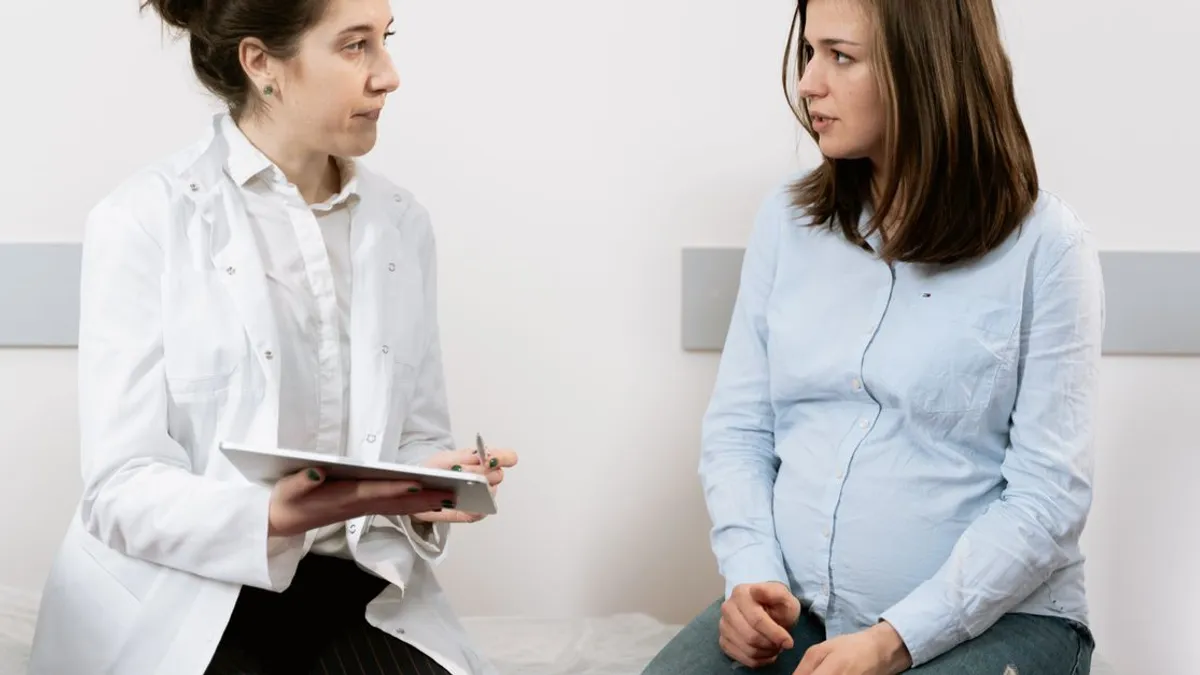
When a patient first hears the words "you have a thyroid nodule", the first reaction is fear, and the question that follows almost automatically is "do I need a biopsy?". A common mistake in general practice is to answer "yes" simply because the nodule exists. In reality, thyroid nodules are an extremely common population finding. Guth et al. (PMID 19601965) showed on high-frequency ultrasound screening that nodules are present in 48–67% of the adult population, and in cohorts over 60 the prevalence exceeds 70%. Clinically significant cancer among these nodules accounts for less than 5%.
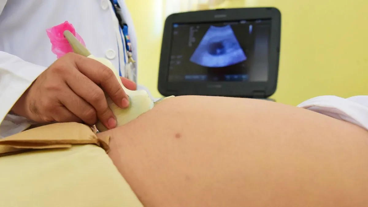
Modern thyroidology resolves this asymmetry with standardized ultrasound risk-stratification scales. The ACR TIRADS (Thyroid Imaging Reporting and Data System), published by Tessler et al. in 2017 (PMID 28372962), and the 2015 American Thyroid Association (ATA) guideline edited by Haugen et al. (PMID 26462967) are the two anchor documents that define who gets FNA and who is followed.
The thesis of this review: a nodule by itself is not an indication for biopsy. The decision is made by the combination of ultrasound appearance (the TR category) and size. TIRADS is the tool that prevents overdiagnosis, unnecessary procedures, and lifelong anxiety in patients with benign findings.
🌀
What TIRADS measures
TIRADS is a standardized scoring system that grades a nodule on five ultrasound features, each assigned a point value. The total score determines the TR-1…TR-5 category and the corresponding malignancy risk.
▸Composition — solid or almost completely solid (2 points), mixed cystic and solid (1), entirely cystic or spongiform (0). Purely cystic nodules are almost never malignant. ▸Echogenicity — hypoechoic (2 points), iso- or hyperechoic (1), very hypoechoic (3), anechoic (0). ▸Shape — taller-than-wide on transverse view, where the anteroposterior dimension exceeds the transverse (3 points, a classic suspicious feature). ▸Margin — smooth or ill-defined (0), lobulated or irregular (2), extra-thyroidal extension (3, a direct sign of invasion). ▸Echogenic foci — none or comet-tail artifacts (0), macrocalcifications (1), peripheral rim calcifications (2), punctate echogenic foci / microcalcifications (3).
The sum yields the TR category: TR-1 (0 points), TR-2 (2 points), TR-3 (3 points), TR-4 (4–6 points), TR-5 (≥7 points). Each category carries an empirically established cancer risk.
🌀
TIRADS categories and cancer risk
Risk stratification under ACR TIRADS is the basis of clinical decision-making. The figures are drawn from the validation cohorts of Hoang et al. (PMID 29498593) and confirmed by subsequent meta-analyses.
▸TR-1 — benign, cancer risk < 1%. Usually entirely cystic nodules or spongiform structure. ▸TR-2 — not suspicious, cancer risk < 2%. Simple cysts, small isoechoic nodules with smooth margins. ▸TR-3 — mildly suspicious, risk ~5%. Isoechoic solid nodule without additional suspicious features. ▸TR-4 — moderately suspicious, risk 5–20%. Hypoechoic plus one or two additional features. ▸TR-5 — highly suspicious, risk > 20%, reaching 35–50% in some cohorts. Markedly hypoechoic plus microcalcifications plus taller-than-wide shape plus lobulated margin.
These numbers are not a theoretical abstraction. They are the basis for the size thresholds at which FNA biopsy is indicated. The higher the TR category, the smaller the size that already requires sampling.
| Category | Cancer risk | FNA threshold |
|---|---|---|
| TR-1 | < 1% | surveillance |
| TR-2 | < 2% | surveillance |
| TR-3 | ~5% | ≥ 2.5 cm |
| TR-4 | 5–20% | ≥ 1.5 cm |
| TR-5 | > 20% | ≥ 1.0 cm |
🌀
When FNA biopsy is indicated
Fine-needle aspiration (FNA) is the standard for cytological verification. Under ACR TIRADS the size thresholds for FNA are:
▸TR-3 at size ≥ 2.5 cm — low risk, biopsy only at large size. ▸TR-4 at size ≥ 1.5 cm — moderate risk, lower threshold. ▸TR-5 at size ≥ 1.0 cm — high risk, biopsy at any nodule ≥ 1 cm. ▸Any nodule with extra-thyroidal extension — FNA required regardless of size. ▸Suspicious cervical lymph nodes (rounded shape, loss of hilum, microcalcifications, peripheral vascularity) — FNA of both the nodule and the lymph node.
An alternative system — the 2015 ATA guideline (PMID 26462967) — operates on five ultrasound risk patterns (benign, very low, low, intermediate, high suspicion). Its size thresholds are similar but not identical to TIRADS. In real practice the endocrinologist uses whichever system the local ultrasound center reports in; what matters is a single standardized description, not a free-text "1.2 cm nodule with heterogeneous structure".
Related article — T4 to T3 conversion and functional hypothyroidism — covers the functional evaluation of the thyroid that runs alongside structural assessment.
🌀
Common pitfalls
General-practice management often errs in two opposite directions. The first is biopsy of any nodule ≥ 1 cm regardless of ultrasound features. The second is declining FNA for a "small" 7–9 mm nodule that under TR-5 already formally requires sampling.
▸Size alone does not decide. Up to 50% of the population has nodules on ultrasound (Guth, PMID 19601965). The vast majority are benign. A nodule > 1 cm without suspicious features is not an indication for biopsy. Conversely, an 8 mm nodule with TR-5 features requires biopsy. ▸Cervical lymph nodes are underassessed. Metastatic involvement of the central (level VI) or lateral (II–V) cervical groups is an independent marker of an aggressive phenotype. Neck ultrasound should be a routine part of nodule evaluation. ▸Extra-thyroidal extension is often not described. It is a direct sign of T3 stage and determines surgical strategy. ▸Thyroid function is ignored at the imaging stage. A "hot" nodule on scintigraphy (autonomous adenoma) almost never undergoes malignant transformation and does not require FNA, but it does require TSH, free T3, free T4, and TRAb assessment. ▸Patient context is not factored in. Childhood head and neck radiation, family history of medullary thyroid cancer (MEN-2), rapid nodule growth, fixation to surrounding tissues, and recurrent laryngeal nerve palsy each lower the biopsy threshold independently of the formal TIRADS criteria.
🌀
Full ultrasound checklist
A high-quality nodule description must contain a standardized set of features. If the ultrasound report states only "1.2 cm nodule with heterogeneous structure", it is insufficient for TIRADS-based decision-making, and a repeat study is required.
▸Composition — solid / cystic / mixed / spongiform. The proportion of solid component is described in mixed nodules. ▸Echogenicity — hypo-, iso-, hyper-, very hypoechoic (relative to strap muscles), anechoic. ▸Shape — anteroposterior to transverse ratio on the axial plane. Taller-than-wide is the classic suspicious feature. ▸Margin — smooth / lobulated / spiculated / extra-thyroidal extension. ▸Calcifications — microcalcifications (punctate, up to 1 mm), macrocalcifications (> 1 mm, often benign when central), peripheral rim (often benign). ▸Doppler vascularity — absent, peripheral, intranodular. Intranodular hypervascularity combined with other suspicious features raises risk. ▸Size in three dimensions — anteroposterior, transverse, longitudinal. ▸Location — right / left / isthmus, upper / mid / lower pole. ▸Regional lymph node status — a mandatory section of the report.
A standardized reporting template (such as the ACR TIRADS template) lets any endocrinologist read the report unambiguously, compare nodules over time, and decide without sending the patient back for repeat imaging by another specialist.
🌀
Surveillance protocol
If the nodule does not meet biopsy threshold, that does not mean "forget about it until symptoms appear". The standard surveillance protocol under ACR TIRADS and ATA:
▸TR-1 / TR-2 — ultrasound in 12 months, then by trajectory. For stable cysts annual or less frequent ultrasound is acceptable. ▸TR-3 < 2.5 cm — ultrasound in 12–24 months. ▸TR-4 < 1.5 cm — ultrasound in 12 months. ▸TR-5 < 1.0 cm — ultrasound in 6–12 months, a more frequent interval due to elevated risk. ▸Stable size for 5 years — interval may be reduced to 24 months. A truly stable nodule is almost always benign. ▸Significant growth criteria — nodule volume increase ≥ 50% (or 20% in two of three dimensions). On significant growth, reassess TIRADS and decide on FNA.
Alongside ultrasound surveillance, TSH, free T3, free T4, and TPO-Ab are assessed every 12 months — to monitor function and detect any autoimmune background that may alter the interpretation of the nodular structure. Related article — low-dose naltrexone (LDN) for thyroid and Hashimoto — covers immunomodulatory strategy in concurrent autoimmune thyroiditis.
🌀
After biopsy — the Bethesda system
FNA cytology is reported using the Bethesda System for Reporting Thyroid Cytopathology — a standardized six-category scale, each with its own malignancy risk and algorithm (Cibas and Ali 2017, PMID 29091573).
▸Bethesda I — non-diagnostic / unsatisfactory (cancer risk 5–10%). Repeat FNA under ultrasound guidance in 3 months. ▸Bethesda II — benign (cancer risk < 3%). Annual ultrasound. No surgery. ▸Bethesda III — atypia of undetermined significance (AUS/FLUS) (cancer risk 10–30%). Repeat FNA or molecular testing (Afirma GSC, ThyroSeq, ThyGeNEXT/ThyraMIR). ▸Bethesda IV — follicular neoplasm / suspicious for follicular neoplasm (cancer risk 25–40%). Molecular testing, or diagnostic lobectomy. ▸Bethesda V — suspicious for malignancy (cancer risk 50–75%). Surgery (thyroidectomy or lobectomy). ▸Bethesda VI — malignant (risk > 99%). Surgery is standard.
Molecular testing in Bethesda III–IV is a growing area. Tests with a high negative predictive value (NPV > 95%, like Afirma GSC) can spare 40–60% of patients with indeterminate cytology from diagnostic surgery. Access varies by region; in the US and EU these tests are standard.
🌀
Monitoring and dynamic follow-up
After the decision on the nodule — biopsy or surveillance — the patient enters dynamic follow-up. Objective evaluation points:
▸Thyroid and neck ultrasound — interval based on TR category (see above). TIRADS-format description is mandatory. ▸TSH, free T3, free T4 — every 6–12 months. Hyperthyroidism in the presence of a nodule requires I-123 scintigraphy to rule out an autonomous adenoma. ▸TPO-Ab, Tg-Ab — always at baseline; repeat if autoimmune thyroiditis is suspected. ▸Calcitonin — if medullary thyroid cancer is suspected (family history of MEN-2, RET mutations) or with suspicious cytology. ▸Thyroglobulin — NOT a marker for benign nodules. Used only after thyroidectomy to monitor for recurrence of differentiated cancer.
Related article — iodine and thyroid: a five-step protocol — covers iodine status assessment, which affects thyroid structure and function independently of the nodular picture.
🌀
Caution — limitations and red flags
TIRADS is a statistical stratification tool, not absolute truth. There are clinical situations where the formal TR class may underestimate risk, and the decision is made individually.
▸Childhood external head and neck irradiation — lower the FNA threshold by one TR category. ▸Family history of medullary or papillary thyroid cancer, MEN-2A/2B, Cowden syndrome, Gardner syndrome, Carney complex — genetic counseling, lower biopsy thresholds. ▸Rapid nodule growth over 6–12 months — an independent feature requiring FNA even at a formally low TR category. ▸Symptoms of invasion — dysphagia, hoarseness, recurrent laryngeal nerve palsy, fixation of the nodule to surrounding tissues — immediate FNA and referral to a thyroid surgeon. ▸Pregnancy — FNA is safe in the second trimester when indicated. Surgery is deferred until after delivery in the absence of rapid growth. ▸Children and adolescents — FNA thresholds are lower than in adults; even a 0.5 cm nodule with suspicious ultrasound features warrants discussion. ▸Multinodular goiter — each nodule is assessed separately under TIRADS; "one large nodule is benign — so the others must be" is not a valid inference. ▸Purely cystic nodule with rapid fluid reaccumulation — even without a solid component, papillary cancer with cystic degeneration is possible. Ultrasound-guided aspiration with cytological analysis of the fluid is warranted.
🌀
Bottom line
A thyroid nodule is not a sentence — it is a question that requires structured assessment. The key rules:
▸The biopsy decision is made by the combination of TR category and size, not by the mere fact of detection. ▸TR-1 / TR-2 — surveillance. TR-3 — biopsy at ≥ 2.5 cm. TR-4 — at ≥ 1.5 cm. TR-5 — at ≥ 1.0 cm. Suspicious lymph nodes and extra-thyroidal extension — biopsy regardless of size. ▸High-quality ultrasound description in a standardized TIRADS format is the obligatory baseline; "heterogeneous structure" is not enough. ▸After FNA, the Bethesda system drives the next step — surveillance, repeat FNA, molecular test, or surgery. ▸Dynamic follow-up is an active strategy, not passive waiting: ultrasound + thyroid function + autoimmune markers per protocol. ▸Patient context (radiation history, family history, growth, symptoms) can lower the biopsy threshold independent of the formal TR category.
TIRADS is the tool that prevents two errors at once: overdiagnosis in 95% of patients with benign nodules and missed aggressive cancer in the small fraction of suspicious cases. Its application requires a trained ultrasound specialist, a clinical endocrinologist, and — in cytologically indeterminate cases — access to molecular testing.
🌀
About the author
I am Dr. Vladimir Pereligyn, endocrinologist and researcher. I specialize in endocrine, metabolic, and autoimmune protocols with a holistic approach and individualized lab diagnostics. Book a consultation — universum.earth/consultation. Daily clinical breakdowns — @md_pereligyn_thyroid.
🌀
Sources
▸Tessler FN, Middleton WD, Grant EG, et al. ACR Thyroid Imaging, Reporting and Data System (TI-RADS): White Paper of the ACR TI-RADS Committee. J Am Coll Radiol. 2017;14(5):587-595. (PMID 28372962) ▸Haugen BR, Alexander EK, Bible KC, et al. 2015 American Thyroid Association Management Guidelines for Adult Patients with Thyroid Nodules and Differentiated Thyroid Cancer. Thyroid. 2016;26(1):1-133. (PMID 26462967) ▸Guth S, Theune U, Aberle J, et al. Very high prevalence of thyroid nodules detected by high frequency (13 MHz) ultrasound examination. Eur J Clin Invest. 2009;39(8):699-706. (PMID 19601965) ▸Hoang JK, Middleton WD, Farjat AE, et al. Reduction in Thyroid Nodule Biopsies and Improved Accuracy with American College of Radiology Thyroid Imaging Reporting and Data System. Radiology. 2018;287(1):185-193. (PMID 29498593) ▸Cibas ES, Ali SZ. The 2017 Bethesda System for Reporting Thyroid Cytopathology. Thyroid. 2017;27(11):1341-1346. (PMID 29091573)
*This article is for informational purposes only and does not replace a medical consultation. Before starting any supplements, changing medication, or undergoing diagnostic procedures, discuss the plan with your physician.*
References
- PMID 19601965. PMID 19601965
- PMID 28372962. PMID 28372962
- PMID 26462967. PMID 26462967
- PMID 29498593. PMID 29498593
- Cibas and Ali 2017, PMID 29091573. PMID 29091573