Introduction: the pain is real, the diagnosis is missing
This story repeats in consulting rooms for years. A woman of 35–45, two births. A dull lower-abdominal pain — not sharp, not crampy, just a constant heaviness that grows louder by evening. The gynecologist checks the ultrasound: "everything is normal." Labs are quiet. Next stop — "it's stress", or "adhesions", or an antidepressant.
Yet the pain has a distinct signature nobody asked about: almost absent in the morning, building through the day, worse with prolonged standing, better lying down, and after intimacy it aches dully for hours. That is not the signature of inflammation, nor of endometriosis. It is the signature of incompetent veins.
Per the 2026 review in the Journal of Clinical Medicine (PMID 41753340[1]), chronic pelvic pain — pain lasting over six months — affects more than 40% of women worldwide and accounts for about 10% of all gynecological visits. A substantial share of these cases, after laparoscopies and courses of "inflammation" treatment, turns out to be venous congestion that was visible from the start — had the veins been examined standing.
Mechanism: hydraulics versus valves
The pelvic veins are a low-pressure valved system working against gravity. In women it faces two unique loads.
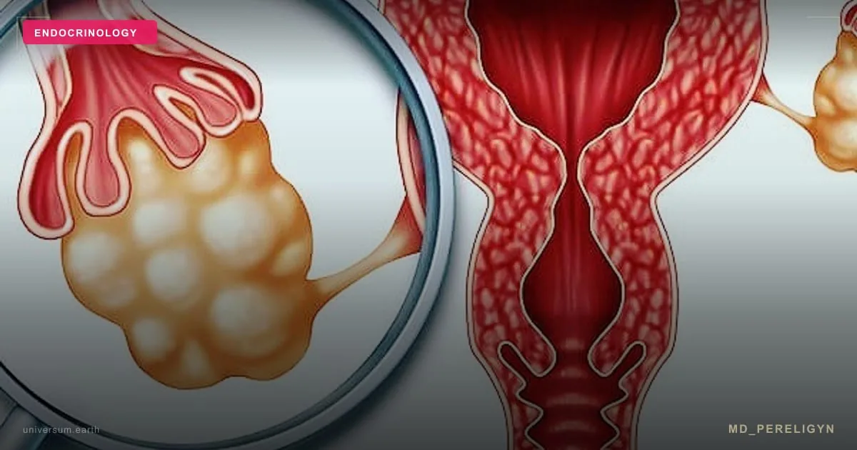
When the ovarian vein valves fail to close, blood flows downward under gravity — reflux. The venous plexuses around the uterus and ovaries engorge, stretch, and turn into slowly emptying reservoirs. A stretched vein does not hurt by itself — pain comes from stretch receptors in the wall and surrounding tissue. Hence the entire clinical picture: the longer the upright posture, the fuller the reservoirs, the stronger the pain.
The symptom profile: three questions that change the diagnosis
The consensus definition (VEIN-TERM, PMID 25873121[2]) describes the syndrome as chronic symptoms that may include pelvic pain, perineal heaviness, urinary urgency and post-coital pain, caused by reflux and/or obstruction of ovarian or pelvic veins, possibly associated with vulvar, perineal and lower-limb varices.
In practice, three questions most often decide the diagnosis.
First: is the pain worse in the morning or in the evening? Venous pain obeys gravity: in the morning, after a night lying down, the reservoirs are empty and pain is nearly absent; by evening, after a day on one's feet, it peaks. Prolonged standing and sitting worsen it; lying with legs elevated relieves it. Inflammatory and endometriosis pain do not behave this way.
Second: what happens after intimacy? A dull ache appearing after intercourse and lasting 30 minutes to a day is the symptom the 2026 review calls pathognomonic for venous congestion. The mechanism is hydraulic: arousal engorges the pelvic plexuses, and incompetent veins take hours to empty. Dyspareunia — pain with deep penetration — occurs in 71–78% of patients. Women almost never volunteer this symptom; it must be asked about.
Third: are there varicose veins where they "should not be"? Veins of the vulva, perineum, buttocks and posterior thigh are the escape routes of pelvic reflux. Varices in these zones — especially appearing in pregnancy and persisting after delivery — are the visible tip of the pelvic problem.
The picture is completed by urinary symptoms — daytime frequency, nocturia and a sensation of incomplete voiding occur in up to 65% of patients per the review (engorged plexuses neighbor the bladder) — and evening perineal heaviness.
| Sign | Venous congestion | Endometriosis | Chronic cystitis |
|---|---|---|---|
| Daily rhythm | absent in the morning, peaks by evening | cycle-bound | tied to voiding |
| Orthostatic effect | worse standing, better lying | independent | independent |
| After intimacy | dull ache 30 min — a day | pain during intercourse | burning, urgency |
| Cyclicity | weak, worse premenstrually | pronounced | none |
| Visible varices | vulva, perineum, thighs | none | none |
🌀
Why the ultrasound is "normal": the physics of body position
The main reason for years of underdiagnosis is banal: a standard pelvic ultrasound is performed supine. Horizontal position removes the gravitational load, the reservoirs partially collapse — and vein diameters return to formally decent values. The physician honestly writes "no pathology", because at the moment of the exam none is visible.
The correct protocol is Doppler examination of the pelvic veins standing or in anti-Trendelenburg position (head down), with a Valsalva maneuver (straining that raises intra-abdominal pressure and provokes reflux). Quantitative criteria per the 2026 review (PMID 41753340[1]):
▸ ovarian vein diameter >7–8 mm (a >8 mm cut-off yields sensitivity comparable to venography); ▸ reflux — reverse flow — lasting >1 second; ▸ low flow velocity <3 cm/s; ▸ arcuate myometrial vein dilatation >5 mm.
The combination of four parameters yields 100% sensitivity; even a single positive parameter — 94.5%. The diagnostic tool is neither exotic nor expensive — it is the same ultrasound, performed in the right position with the right maneuver. When in doubt, the next step is MR venography, and catheter venography remains the gold standard, usually combined with treatment.
The practical takeaway for the patient: if your ultrasound report does not mention body position and Valsalva — venous congestion has not been excluded. It was not looked for.
Left-sided pain: two compression syndromes
An anatomical detail explains why pelvic congestion is more often left-sided: the left ovarian vein drains not directly into the inferior vena cava, like the right, but into the left renal vein — at a right angle and with a longer path against gravity.
Two scenarios deserve separate attention. Nutcracker syndrome: the left renal vein is compressed between the aorta and the superior mesenteric artery; pressure rises and pushes blood down the ovarian vein. A clue is microhematuria on urinalysis. May–Thurner syndrome: the left common iliac vein is compressed by the right iliac artery; here the problem is outflow obstruction, not reflux.
Distinguishing these scenarios is not academic — it determines treatment: embolization with undiagnosed compression will not solve the problem, because the cause of stasis is upstream. A 2026 systematic review (PMID 42372331[3]) separately notes that pelvic venous obstruction can also manifest as orthostatic intolerance — palpitations and lightheadedness when upright — due to blood pooling below the diaphragm.
Treatment: what is proven and what is not
The hierarchy by invasiveness and by state of the evidence.
Venoactive drugs (micronized purified flavonoid fraction and other flavonoids). The Phlebology 2026 systematic review (PMID 41432346[4]) rates the evidence in venous pelvic pain as limited: small heterogeneous studies, a possible symptomatic effect, no large high-quality RCT. A 2025 pilot (PMID 40049814[5]) showed reduced venous diameter and improved quality of life, but it is an unblinded pilot. The honest framing: a reasonable low-risk first step, not a cure.
Compression and behavioral measures — avoiding hours of static standing, breaks lying with legs elevated, treating constipation (straining is a chronic reflux provocateur). Modest evidence, direct mechanistic logic, zero risk.
Ovarian vein embolization — catheter occlusion of refluxing veins with coils and/or sclerosant via an arm or groin vein puncture, no incisions. A 2025 before-and-after quality-of-life study (PMC12517425) showed significant pain reduction and questionnaire improvement after the procedure. New techniques are emerging — endovenous thermal ablation of pelvic veins (PMID 41519470[6]), so far at the descriptive-series level.
Iliac vein stenting — for obstructive forms (May–Thurner). A long-follow-up series (PMID 39401615[7]) demonstrates durable multi-year relief of chronic pelvic pain in properly selected patients with confirmed obstruction.
The general selection principle is formulated in a 2026 paper on treatment sequencing (PMID 42526747[8]): treat the dominant hemodynamic lesion confirmed on imaging — embolize reflux, stent obstruction, not the other way around.
The endocrine angle: where venous congestion meets hormones
Why this topic sits in my library next to the thyroid. Three intersections.
What is NOT proven
▸Venoactive drugs as a cure. Limited data allow a symptomatic effect; a tablet does not fix the structural valve problem. ▸A one-to-one link between vein size and pain intensity. Dilated pelvic veins are also found in asymptomatic women; one treats a symptomatic patient with confirmed hemodynamics, not an image. ▸An effect of venous congestion on the hormonal profile. Mechanistic conjectures exist, quality clinical data do not — I do not use this claim. ▸Embolization as a guarantee. Quality-of-life improvement is shown, but predictors of non-response are poorly studied; part of the pain may have a second source (coexisting endometriosis, a myofascial component).
Conclusions
Pelvic congestion syndrome has a clear clinical signature: dull non-cyclical pain lasting over six months, building toward evening and with standing, relieved lying down, pathognomonic post-coital ache, vulvar and perineal varices after pregnancies, urinary symptoms without cystitis.
The diagnosis requires no exotics — it requires a properly performed Doppler: standing or anti-Trendelenburg, with Valsalva, with numbers in the report (diameter >7–8 mm, reflux >1 s, flow <3 cm/s). Treatment follows the confirmed hemodynamics: reflux — embolization, obstruction — stenting, mild forms — the conservative tier.
If you recognized your pain in this description — start with the three questions from this article and ask for a referral for pelvic vein Doppler with a Valsalva maneuver. It is one visit that can end the years-long loop of "ultrasound is normal — it's stress".
This article is for information purposes and does not replace a medical consultation. Pelvic congestion syndrome is diagnosed from the combination of clinical findings and imaging; treatment decisions are made by a physician after evaluation.
Sources
▸Krambeck C, et al. Pelvic Congestion Syndrome: The Gynecological Perspective. J Clin Med 2026. PMID 41753340[1], PMC12941841 — review; source of symptom frequencies (dyspareunia 71–78%, urinary symptoms up to 65%, dilatation in >63% multiparous vs 10%) and ultrasound criteria (>7–8 mm, reflux >1 s, <3 cm/s, 100% sensitivity of the combination). ▸Gavorník P, et al. Pelvic venous congestion syndrome — diagnosis and management (VEIN-TERM definition). Vnitr Lek 2015. PMID 25873121[2] — consensus definition. ▸Marcelin C, et al. Diagnosis and Management of Pelvic Venous Disorders in Females. Diagnostics 2022. PMID 36292025[9], PMC9600975 — diagnostic and management review. ▸Durham JD, Machan L. Pelvic congestion syndrome. Semin Intervent Radiol 2014. PMID 24436564[10], PMC3835435 — classic review. ▸Bałabuszek K, et al. Symptoms and quality of life assessment after coil and foam embolization in patients with venous-origin chronic pelvic pain. Ann Med 2025. PMID 41074660[11], PMC12517425 — quality of life before/after embolization. ▸Gloviczki ML, et al. Venoactive drugs for venous origin chronic pelvic pain in women: a systematic review. Phlebology 2026. PMID 41432346[4] — evidence for venoactive drugs is limited. ▸Lee IH, et al. Medical treatment for pelvic congestion syndrome with flavonoid: a pilot study. Taiwan J Obstet Gynecol 2025. PMID 40049814[5] — flavonoid pilot. ▸Villalba L, Larkin T. Iliac venous stenting provides long-term relief from chronic pelvic pain. J Vasc Surg Venous Lymphat Disord 2025. PMID 39401615[7] — stenting for obstruction. ▸Li X, et al. Outcomes of symptom-driven top-down versus bottom-up strategies for pelvic-origin extra-pelvic varicose veins. Ann Vasc Surg 2026. PMID 42526747[8] — treatment sequencing principle. ▸Moreno L, et al. A novel endovascular thermal ablation technique for pelvic venous disorders. J Vasc Surg Venous Lymphat Disord 2026. PMID 41519470[6] — new technique, descriptive series. ▸Imami MR, et al. Pelvic venous disorders and orthostatic intolerance: a systematic review. Auton Neurosci 2026. PMID 42372331[3] — pelvic venous obstruction and orthostatic intolerance.
Key facts
- Chronic pelvic pain — pain lasting longer than 6 months — affects over 40% of women worldwide per a 2026 J Clin Med review and accounts for about 10% of gynecological consultations.
- The consensus definition of pelvic congestion syndrome (VEIN-TERM): chronic pain + perineal heaviness + urinary urgency + post-coital pain, caused by reflux and/or obstruction of ovarian or pelvic veins.
- Dull post-coital pain lasting 30 minutes to a day is called pathognomonic in the 2026 review; dyspareunia occurs in 71–78% of patients.
- Pelvic venous dilatation is observed in over 63% of multiparous women versus 10% of nulliparous — pregnancy is the leading risk factor.
- The key venous pain pattern: worse by evening and with prolonged standing, relieved by lying down and almost absent in the morning — inflammatory and endometriosis pain do not behave this way.
- Ultrasound criteria: ovarian vein diameter >7–8 mm, reflux >1 s on Valsalva, flow velocity <3 cm/s, arcuate myometrial vein dilatation >5 mm; the combination of four parameters yields 100% sensitivity.
- A standard supine pelvic ultrasound misses the diagnosis: the exam must be performed standing or in anti-Trendelenburg position with a Valsalva maneuver.
- Ovarian vein embolization improves quality of life in venous pelvic pain (PMC12517425); iliac vein stenting for obstruction provides long-term relief (PMID 39401615); a 2026 systematic review rates the evidence for venoactive drugs as limited.
- Left-sided pain warrants exclusion of compression syndromes: nutcracker (aortomesenteric) and May–Thurner.