Introduction: the price of accuracy is radiation exposure
Myocardial perfusion scintigraphy (SPECT, single-photon emission computed tomography) with technetium-99m remained the workhorse for ischemia assessment for decades. The method provides a quantitative perfusion map, identifies areas of reduced blood flow, and helps determine whether revascularization is appropriate.
The price of accuracy is a radiation dose of ~12 mSv (millisieverts) per examination. This is equivalent to approximately 600 chest X-rays or four times the annual background radiation exposure. In a patient who undergoes repeated SPECT studies, the cumulative dose over a decade may reach 50–60 mSv, a statistically significant increase in oncologic risk (Berrington de González A, Lancet 2004, PMID 14738798[1]).
Key idea of the md_pereligyn protocol: by 2026, a cardiologist has a set of noninvasive modalities that provide comparable or greater diagnostic accuracy without ionizing radiation. A request for “SPECT” is justified only when the first three alternatives cannot be performed.
This article explains what works instead of SPECT, which modality is indicated for whom, and what modern cardiac screening looks like in my practice.
🌀
The dose of one SPECT equals 600 chest X-rays
12 mSv for one test is not a “small scan.” It is the annual occupational background radiation limit for a healthcare worker accumulated in half an hour. Comparisons for perspective:
▸1 chest X-ray — 0.02 mSv ▸1 chest CT (standard protocol) — 7 mSv ▸1 coronary calcium CT (CAC-score) — 1 mSv ▸1 myocardial SPECT — 12 mSv ▸1 whole-body PET-CT (FDG) — 25 mSv ▸Annual natural background — 2.4 mSv ▸Annual public dose limit (ICRP) — 1 mSv
The linear no-threshold model of radiation risk assumes that each additional mSv adds a small but measurable increase in lifetime oncologic risk. In younger patients and women, this risk is higher because of greater tissue radiosensitivity, especially breast tissue during chest imaging.
This is not a reason to never perform SPECT. It is a reason not to use it routinely when alternatives provide the same or more information.
🌀
Alternatives: what and for whom
### 1. CAC-score (coronary calcium) — for asymptomatic screening
Coronary calcium CT is a low-dose non-contrast cardiac CT. The radiation dose is ~1 mSv, 12 times lower than SPECT. It directly visualizes atherosclerotic plaque as calcifications in the coronary arteries.
▸Principle: atherosclerotic plaque calcifies during fibrotic stabilization. The more calcium there is in the coronary arteries, the greater the total atherosclerotic burden. ▸Agatston scale: - 0 units — no atherosclerosis, very low risk (10-year CHD risk <1%) - 1–10 — minimal - 11–100 — moderate - 101–400 — high - >400 — very high (event risk 10 times higher than baseline) ▸Prognostic value: in asymptomatic patients aged 40+, CAC-score outperforms traditional risk scales (Framingham, ASCVD). The MESA study showed that CAC = 0 reclassifies patients from “intermediate risk” to “low risk” with a 10-year risk <2% (Greenland P, JACC 2010, PMID 20428003[2]). ▸DETRANO 2008: CAC > 300 increases the risk of coronary events 9.7-fold compared with CAC = 0 (Detrano R, NEJM 2008, PMID 18367736[3]).
Who it is indicated for: ▸Asymptomatic patients aged 40+ with one risk factor (family history, hypertension, diabetes, smoking) ▸Before prescribing a statin for primary prevention in patients with borderline lipids ▸After any “normal” standard screening when there is uncertainty about true risk
### 2. Stress echocardiography — for women and suspected ischemia
Cardiac ultrasound under physical or pharmacologic (dobutamine) stress. 0 mSv radiation. It detects regional ventricular wall motion abnormalities during induced ischemia.
▸Sensitivity 80–85%, specificity 84–86% — comparable to SPECT (Geleijnse ML, Eur Heart J 2009, PMID 19297421[4]). ▸Especially valuable in women — no breast irradiation and no breast attenuation artifacts, which often reduce SPECT quality. ▸Dynamic assessment — shows the functional behavior of the myocardium in real time, not only a static perfusion map. ▸Additional parameters: global longitudinal strain (GLS), diastolic function, pulmonary artery pressure, valve status.
Who it is indicated for: ▸Clinical suspicion of ischemia in women (first-line choice) ▸Atypical chest pain with preserved ejection fraction ▸Assessment of functional reserve after revascularization ▸Risk stratification before noncoronary surgery
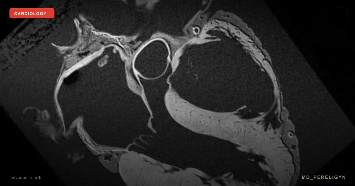
### 3. CMR (cardiac MRI) — the gold standard for viability
Cardiac magnetic resonance with a perfusion protocol and gadolinium contrast. 0 mSv ionizing radiation. Extremely high soft-tissue resolution.
▸Myocardial viability — late gadolinium enhancement (LGE) accurately distinguishes viable myocardium from scar. This is critical before deciding on revascularization: scar tissue does not respond to revascularization. ▸Subendocardial ischemia — CMR detects a thin layer of endocardial ischemia that SPECT cannot see. ▸T1/T2 mapping — quantitative assessment of edema, fibrosis, amyloidosis, and myocarditis. ▸Accurate ejection fraction — the gold standard, independent of Simpson geometric assumptions as in echocardiography.
Who it is indicated for: ▸Post-infarction viability assessment before coronary revascularization ▸Suspected myocarditis, amyloidosis, hypertrophic cardiomyopathy ▸Atypical cardiomyopathies of unclear etiology ▸Family history of sudden cardiac death (to exclude arrhythmogenic right ventricular cardiomyopathy)
### 4. HRV (heart rate variability) — functional screening of the autonomic nervous system
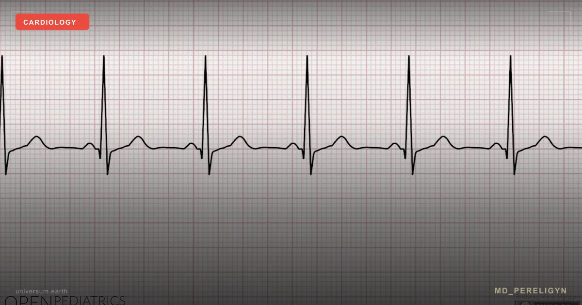
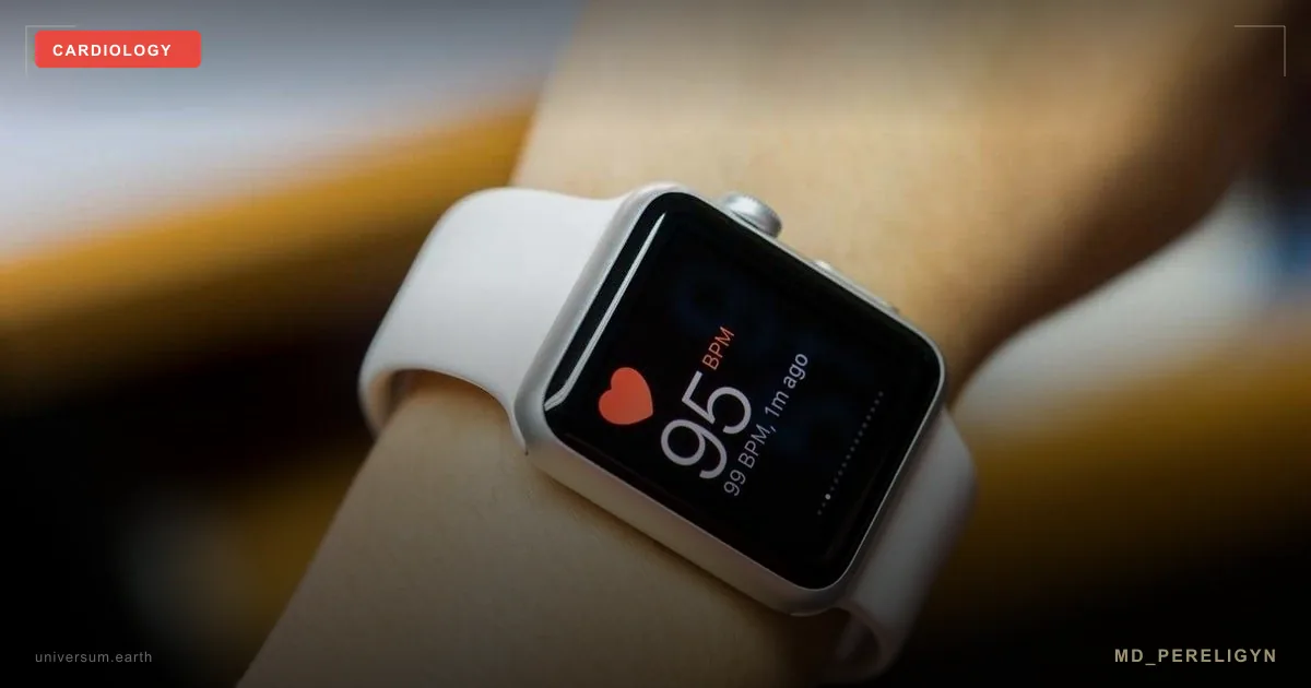
Heart rate variability is a measure of differences in R-R intervals between heartbeats. It reflects the balance between the sympathetic and parasympathetic nervous systems. 0 mSv, noninvasive, and available through any modern wearable ECG sensor (Polar H10, Apple Watch, Whoop, Oura).
▸RMSSD (root mean square of successive differences) — the main marker of parasympathetic tone. Normal range for adults is 20–60 ms. ▸SDNN (standard deviation of NN intervals) — overall variability over 24 hours. Normal >100 ms. ▸LF/HF ratio — the ratio of sympathetic to parasympathetic contribution. Normal 1.5–2.0. ▸Low HRV is associated with a twofold increase in the risk of sudden cardiac death in post-infarction patients (Tsuji H, Circulation 1996, PMID 8902162[5]). ▸A decrease in HRV often precedes clinical symptoms of coronary artery disease, chronic stress, and autonomic dysfunction in diabetes.
Who it is indicated for: ▸Screening for autonomic dysfunction in patients with diabetes (diabetic cardiac neuropathy) ▸Assessment of recovery after exercise in athletes ▸Monitoring the effectiveness of stress management in a holistic program ▸Prognostic marker in post-infarction patients
### 5. VO2max — the gold standard for cardiorespiratory fitness
Maximal oxygen uptake during incremental exercise (cycle ergometer or treadmill with gas analysis). Measured in mL/kg/min. It is a stronger prognostic marker than blood pressure, LDL-C, and smoking combined (Mandsager K, JAMA Netw Open 2018, PMID 30537017[6]).
▸VO2max ≥35 mL/kg/min — low CVD risk ▸VO2max 25–34 — moderate risk ▸VO2max <20 — high CVD risk, with 5-fold higher mortality compared with the “elite” athlete group ▸Each 1 MET (3.5 mL/kg/min) increase in VO2max reduces mortality by 12% (Kodama S, JAMA 2009, PMID 19454641[7]).
Who it is indicated for: ▸Functional reserve screening in any patient aged 40+ ▸Baseline assessment before starting an exercise program ▸Objective evaluation of intervention effect (repeat after 3–6 months) ▸Risk stratification before noncardiac surgery
### 6. FMD (flow-mediated dilation) — endothelial function
Brachial artery ultrasound before and after 5-minute cuff occlusion — measurement of reactive vessel dilation in response to hyperemia. 0 mSv, noninvasive, and the test takes 30 minutes.
▸FMD >7% — healthy endothelial function ▸FMD 4–7% — borderline ▸FMD <4% — pronounced endothelial dysfunction ▸Prognostic value: a 1% decrease in FMD increases the risk of cardiovascular events by 13% (Inaba Y, Int J Cardiovasc Imaging 2010, PMID 20127417[8]). ▸FMD responds to therapy (polyphenols, omega-3, L-arginine) within 8–12 weeks. This is the main objective marker of effectiveness for the holistic protocol.
Who it is indicated for: ▸Screening for preclinical atherosclerosis in patients aged 30+ with risk factors ▸Monitoring the effectiveness of the holistic protocol ▸Selecting patients for the “reversibility” group (FMD 4–7% is the optimal moment for intensive intervention)
🌀
Which test for whom: selection algorithm
▸Asymptomatic screening in patients aged 40+ with risk factors → CAC-score (1 mSv, direct plaque visualization, outperforms risk scales) ▸Clinical suspicion of ischemia, women → stress echocardiography (0 mSv, no breast irradiation) ▸Post-infarction viability assessment → CMR (0 mSv, gold-standard LGE) ▸Functional reserve and exercise strategy → VO2max ▸Effectiveness of the holistic protocol → FMD + omega-3 index + ApoB ▸Autonomic function, diabetic neuropathy → HRV (24-hour Holter or 7-day wearable sensor) ▸Suspected myocarditis, cardiomyopathy, amyloidosis → CMR ▸A request for SPECT is justified only when the first three options cannot be performed, for example because of contrast contraindications, poor ultrasound window, or unavailable CMR
🌀
What does NOT work, or works worse than it seems
▸Standard resting ECG — detects ischemia only during an acute episode or after a prior myocardial infarction. Sensitivity for occult coronary artery disease is <30%. It does not replace any of the methods above. ▸Standard treadmill test (exercise ECG without imaging) — sensitivity 65–70%, specificity 70%. In women, false-positive results reach up to 50%. It is inferior to stress echocardiography across all parameters. ▸Coronary angiography “just in case” — an invasive procedure with a radiation dose of 5–7 mSv and risks including contrast-induced nephropathy, dissection, and bleeding. It is justified only when ischemia has been proven and revascularization is being considered. ▸Isolated CAC-score without assessment of inflammation and lipoproteins — provides the current plaque picture but does not show progression. CAC = 0 does not exclude vulnerable soft plaque. ▸Routine SPECT when alternatives are available — unjustified radiation exposure.
🌀
When to seek evaluation
▸Family history of coronary artery disease, myocardial infarction, or sudden death before age 60 ▸Age 40+ with any risk factor (hypertension, diabetes, smoking, visceral obesity) ▸Atypical chest pain or exertional dyspnea without an obvious cause ▸Before prescribing a statin for primary prevention ▸After “normal” standard screening when you feel that something is wrong ▸A desire to objectively assess the effect of a holistic protocol ▸Before high-risk noncardiac surgery
I perform comprehensive noninvasive cardiac screening (CAC-score, stress echocardiography, FMD, VO2max, HRV, extended lipid profile with ApoB and Lp(a)) and develop a personalized vascular risk reduction protocol without excessive radiation.
🌀
Conclusion
SPECT with technetium-99m is a technology of the 1990s. It remains useful in narrow clinical situations, but routine use for screening and risk stratification in 2026 is not justified.
Modern cardiac screening is built on a combination of CAC-score (1 mSv) + stress echocardiography (0 mSv) + CMR (0 mSv) + FMD + VO2max + HRV + extended lipid profile. This combination provides a more complete, earlier, and more prognostically accurate picture of vascular health than a single SPECT study with a dose of 12 mSv.
Radiation is the price paid when there is no alternative. A modern cardiologist almost always has an alternative.
🌀
Sources
▸Greenland P, Bonow RO, Brundage BH, et al. ACCF/AHA clinical expert consensus document on coronary artery calcium scoring. *JACC* 2010;55:e1–e10. PMID 20428003 ▸Detrano R, Guerci AD, Carr JJ, et al. Coronary calcium as a predictor of coronary events in four racial or ethnic groups. *NEJM* 2008;358:1336–1345. PMID 18367736 ▸Budoff MJ, Achenbach S, Blumenthal RS, et al. Assessment of coronary artery disease by cardiac CT: AHA scientific statement. *J Am Coll Cardiol* 2007;49:378–402. PMID 17481443 ▸Berrington de González A, Darby S. Risk of cancer from diagnostic X-rays. *Lancet* 2004;363:345–351. PMID 14738798 ▸Geleijnse ML, Krenning BJ, Nemes A, et al. Stress echocardiography. *Eur Heart J* 2009;30:1255–1268. PMID 19297421 ▸Tsuji H, Larson MG, Venditti FJ Jr, et al. Impact of reduced heart rate variability on risk for cardiac events: Framingham. *Circulation* 1996;94:2850–2855. PMID 8902162 ▸Mandsager K, Harb S, Cremer P, et al. Association of cardiorespiratory fitness with long-term mortality. *JAMA Netw Open* 2018;1:e183605. PMID 30537017 ▸Kodama S, Saito K, Tanaka S, et al. Cardiorespiratory fitness as a quantitative predictor of all-cause mortality. *JAMA* 2009;301:2024–2035. PMID 19454641 ▸Inaba Y, Chen JA, Bergmann SR. Prediction of future cardiovascular outcomes by FMD of brachial artery. *Int J Cardiovasc Imaging* 2010;26:631–640. PMID 20127417 ▸Ridker PM, Danielson E, Fonseca FA, et al. Rosuvastatin to prevent vascular events in men and women with elevated CRP. *NEJM* 2008;359:2195–2207. PMID 18997196
Related articles: Endothelium: the foundation of vascular health, Cholesterol without statins.
🌀
FAQ
The cardiologist insists on SPECT. Should I agree? It depends on the clinical situation. If the indication is asymptomatic risk screening or assessment of ischemia probability with preserved function, it is reasonable to start with CAC-score (1 mSv) or stress echocardiography (0 mSv). SPECT is justified if these modalities are unavailable, produced an equivocal result, or are contraindicated. Discuss the specific rationale for 12 mSv of radiation exposure with your physician.
CAC-score = 0 — is this a complete guarantee of safety? No, but almost. CAC = 0 at age 40+ is associated with a 10-year coronary event risk <1%. However, CAC detects only calcified plaque. A young “soft” lipid plaque without calcification, typical of patients under 50 with familial hypercholesterolemia or high Lp(a), may not be reflected. In patients with Lp(a) >75 mg/dL, additional stress echocardiography or CCTA is justified.
How often should CAC-score be repeated? With CAC = 0 at age 40–55, repeat after 5 years, the so-called “warranty period.” With CAC 1–10, repeat after 3–4 years. With CAC >100, repeat testing is not needed; the diagnosis of atherosclerosis is established, and the focus shifts to aggressive control of risk factors. Each repeat adds 1 mSv of radiation exposure, so do not repeat it without clinical necessity.
Which is better for women: SPECT or stress echocardiography? Stress echocardiography. In women, SPECT often produces false-positive results because of breast tissue artifacts and smaller heart size. Stress echocardiography has comparable sensitivity (80–85%) and specificity (84–86%), requires no radiation, and does not irradiate breast tissue, which is a critical factor for women of reproductive age.
Can VO2max and HRV be measured at home? VO2max can be estimated approximately using Cooper tests, the 6-minute walk test, or heart-rate-based estimation formulas (Apple Watch, Garmin, and Polar provide an estimate). Accurate measurement requires laboratory gas analysis. HRV is available through any modern wearable sensor (Apple Watch, Oura, Whoop, Polar H10); home measurements are sufficient for screening. Clinical decisions require 24-hour Holter monitoring.
*This article is informational and does not replace medical consultation. Before starting any nutraceuticals, changing medication therapy, or undergoing diagnostic procedures, discuss the plan with your treating physician.*
🌀
Coronary CT angiography and FFR-CT: the anatomical-functional hybrid
The original test menu omits coronary computed tomography angiography (CCTA), which has become the first-line non-invasive test for stable chest pain in both the 2021 AHA/ACC and 2024 ESC chest-pain guidelines. CCTA visualises the coronary lumen and vessel wall with sub-millimetre spatial resolution after iodinated contrast injection. Effective radiation dose with modern prospective ECG-gated protocols on 64-slice or higher scanners is 1–3 mSv — comparable to a CAC-score and roughly four-fold lower than SPECT PMID: 30571565.
Two large outcome trials anchor the evidence base. PROMISE (10 003 patients with stable chest pain, median follow-up 25 months) compared CCTA-first versus functional-testing-first strategies; the composite endpoint of death, myocardial infarction, hospitalisation for unstable angina, or major procedural complication did not differ between groups, but CCTA detected more obstructive disease and led to earlier statin initiation. SCOT-HEART (4 146 patients, 5-year follow-up) demonstrated a clinically meaningful reduction in fatal and non-fatal myocardial infarction in the CCTA-guided arm versus standard care alone.
The principal limitation of pure anatomical CCTA is moderate specificity for haemodynamically significant stenosis: a 50–70% lumen narrowing on CT may or may not produce ischaemia. FFR-CT (computed fractional flow reserve, derived by computational fluid dynamics from the standard CCTA dataset without additional scanning, contrast, or radiation) closes this gap. An FFR-CT value ≤0.80 identifies vessel-specific ischaemia with sensitivity 86% and specificity 79% versus invasive FFR as reference standard PMID: 26693139. The technology is FDA- and CE-cleared and reimbursed in several European systems.
Practical exclusions for CCTA: heart rate uncontrollable below 65 bpm despite beta-blockade, severe coronary calcification (CAC >1 000 — blooming artefact obscures lumen), atrial fibrillation with rapid response, iodinated-contrast allergy, eGFR <30 ml/min/1.73 m². For patients with CAC 400–1 000, image quality is borderline; CMR stress perfusion or stress echocardiography is preferred.
🌀
Pre-test probability: who actually needs any imaging
Indiscriminate testing of low-probability patients produces a high false-positive rate, downstream invasive angiography without revascularisation, and net harm. The 2024 ESC chest-pain guideline replaced the older Diamond–Forrester model with a recalibrated tool incorporating age, sex, symptom type, and risk factors. A pre-test probability of obstructive coronary artery disease below 5% identifies patients in whom no further testing is warranted — the negative predictive value of clinical assessment alone exceeds 95% in this band PMID: 36017568.
The recalibrated probabilities are substantially lower than historical Diamond–Forrester estimates. For example, a 50-year-old woman with non-anginal chest pain has a pre-test probability of approximately 2%, not the 13% predicted by older models. Treating these patients with imaging adds cost, anxiety, and false positives without improving outcomes.
The decision rule is layered:
- Pre-test probability <5%: defer imaging; address modifiable risk factors and reassess if symptoms change. - Pre-test probability 5–15%: CAC-score is the rational gatekeeper. CAC = 0 reclassifies most of these patients to <5% probability and event rates of 0.5%/year, comparable to age-matched controls without symptoms [PMID: 33933205](https://pubmed.ncbi.nlm.nih.gov/33933205/). - Pre-test probability 15–50%: CCTA is first-line. - Pre-test probability >50%: functional imaging (stress CMR, stress echo, or invasive coronary angiography with FFR) is preferred because the prior probability of needing intervention already justifies it.
This algorithm shifts roughly 30–40% of patients away from any imaging in real-world cohorts without missing clinically relevant disease, and it is the framework against which every alternative test in this article should be selected.
🌀
Contraindications and adverse events: stress echocardiography and CMR
The article identifies stress echo and CMR as preferred radiation-free alternatives but does not describe their safety profile, which patients ask about before scheduling.
Stress echocardiography. Exercise stress echo carries the same cardiovascular risk as standard treadmill testing: mortality approximately 0.5 per 10 000 studies, non-fatal myocardial infarction approximately 3.5 per 10 000 PMID: 22277455. Dobutamine stress echo (used when patients cannot exercise) has a slightly higher adverse-event rate. Dobutamine is infused stepwise at 5, 10, 20, 30, and 40 µg/kg/min; atropine 0.25–1 mg IV is added if target heart rate (85% of age-predicted maximum) is not reached. Major adverse events occur in 0.3% of studies: sustained ventricular tachycardia (0.15%), atrial fibrillation (1.0%), severe hypotension (1.5%), and rarely myocardial infarction (0.02%) PMID: 18387438. Absolute contraindications: acute coronary syndrome within 48 hours, decompensated heart failure, severe aortic stenosis with valve area <1.0 cm², uncontrolled arrhythmia, severe hypertension (>200/110 mmHg at rest), aortic dissection. Esmolol (0.5 mg/kg IV bolus) is the standard reversal agent for dobutamine-induced ischaemia or arrhythmia.
Cardiac magnetic resonance. Stress CMR uses adenosine or regadenoson for vasodilator perfusion imaging rather than exercise. Adverse events with regadenoson 0.4 mg IV bolus: transient dyspnoea (28%), chest discomfort (13%), headache (26%), AV block (rare, <0.1%) PMID: 28122885. Aminophylline 100–200 mg IV reverses regadenoson effects in seconds. Gadolinium-based contrast agents — used in 80% of clinical CMR studies for late gadolinium enhancement — are contraindicated when eGFR is below 30 ml/min/1.73 m² because of nephrogenic systemic fibrosis risk, although modern macrocyclic agents (gadobutrol, gadoterate) have reduced this risk to near zero PMID: 30025725. Absolute MR contraindications remain: non-MR-conditional cardiac implantable electronic devices, ferromagnetic intracranial aneurysm clips, cochlear implants without MR labelling, and metallic intraocular foreign bodies. Severe claustrophobia affects approximately 4% of patients and can usually be managed with short-bore wide systems or oral lorazepam 0.5–1 mg.
🌀
High-sensitivity troponin as a functional biomarker
The article focuses on imaging and physiological tests but omits the most clinically useful blood biomarker in this domain: high-sensitivity cardiac troponin T or I (hs-cTn). Modern fifth-generation assays detect circulating troponin at concentrations of 1–3 ng/L — three orders of magnitude below the 99th percentile upper reference limit of 14 ng/L (hs-cTnT) or 16–34 ng/L (hs-cTnI, sex-specific). Detectable resting troponin in apparently healthy individuals is not analytical noise; it reflects subclinical myocardial injury and tracks long-term cardiovascular mortality independently of CAC-score, LDL-C, and clinical risk scores PMID: 32860505.
In population cohorts, every doubling of hs-cTnT increases the 10-year risk of cardiovascular death by approximately 25%. A baseline hs-cTnT above 5 ng/L in asymptomatic adults aged 50–75 confers a hazard ratio of 2.5 for incident coronary events over 10 years compared with troponin below the limit of detection, independent of traditional risk factors PMID: 32860505.
Clinical use in functional cardiac assessment:
- Symptomatic patients with normal stress imaging but hs-cTn above the 99th percentile should be reassessed — false-negative imaging is more common than a true troponin elevation in this context. - Asymptomatic adults with hs-cTnT 5–14 ng/L have intermediate residual risk and are reasonable candidates for CAC-scoring even when LDL-C and ASCVD scores appear acceptable. - Serial measurement (annual) tracks treatment response: statin initiation reduces hs-cTnT by approximately 10% within 12 months, and ezetimibe adds an incremental reduction; the magnitude of change correlates with subsequent event reduction.
Troponin does not replace imaging — it does not localise disease and does not differentiate ischaemia from non-ischaemic injury such as myocarditis, amyloidosis, or chronic kidney disease. It is, however, the cheapest and most reproducible functional readout available, and it should be measured before any of the imaging modalities discussed above when long-term risk reclassification is the goal rather than acute diagnosis.
References
- PMID 14738798. PMID 14738798
- PMID 20428003. PMID 20428003
- PMID 18367736. PMID 18367736
- PMID 19297421. PMID 19297421
- PMID 8902162. PMID 8902162
- PMID 30537017. PMID 30537017
- PMID 19454641. PMID 19454641
- PMID 20127417. PMID 20127417
- Budoff MJ, Achenbach S, Blumenthal RS, et al. Assessment of coronary artery disease by cardiac CT: AHA scientific statement. J Am Coll Cardiol 2007;49:378–402. PMID 17481443
- Ridker PM, Danielson E, Fonseca FA, et al. Rosuvastatin to prevent vascular events in men and women with elevated CRP. NEJM 2008;359:2195–2207. PMID 18997196
- PMID: 30571565. PMID 30571565
- PMID: 26693139. PMID 26693139
- PMID: 36017568. PMID 36017568
- PMID: 33933205. PMID 33933205
- PMID: 22277455. PMID 22277455
- PMID: 18387438. PMID 18387438
- PMID: 28122885. PMID 28122885
- PMID: 30025725. PMID 30025725
- PMID: 32860505. PMID 32860505